Implant Pocket Planning
Submuscular, Subglandular and Dual-Plane Breast Augmentation
Compare submuscular, subglandular and dual-plane implant placement through tissue coverage, muscle movement, ptosis, risks and individual selection factors.
The images on this page are conceptual representations; they do not show a real patient, an operation or a treatment result.
When planning breast augmentation, implant volume and shape are not the only decisions. The anatomical level at which the implant will sit must also be considered. The everyday terms “over the muscle”, “under the muscle” and “dual plane” describe the implant’s relationship to the breast gland, pectoralis major muscle and surrounding tissues. Although the labels sound simple, each may cover different surgical details. Implant placement therefore cannot be selected safely from photographs or an online checklist alone.
In brief: In subglandular placement, the implant sits behind the breast tissue and in front of the pectoralis major. “Under the muscle” can describe arrangements in which all or part of the implant lies behind muscle. Dual plane is an individually adjusted partial subpectoral approach in which the upper implant has muscle coverage while the lower breast tissue and muscle–implant relationship are managed differently. There is no universally best plane. Tissue thickness, breast footprint, ptosis, chest wall, muscle activity, implant dimensions and personal priorities must be assessed together.
What does the implant plane mean?
The implant plane is the anatomical position of the pocket created by the surgeon. From superficial to deep, the area includes skin, subcutaneous fat, breast tissue, pectoral fascia, pectoralis major and chest wall. Planning considers not only the names of these layers but their thickness, elasticity and ability to cover an implant in its upper and lower portions.
“Under the muscle” does not always mean that the whole implant is enclosed beneath a single muscle. In several commonly used augmentation techniques, the upper implant is covered by pectoralis major while its lower portion extends below the muscle’s lower border. Ask your surgeon to show the proposed pocket on a drawing; this is usually clearer than hearing a label alone.
What is subglandular, or over-the-muscle, placement?
In a subglandular pocket, the implant is positioned behind the breast gland and in front of pectoralis major. Because the muscle is not elevated over the implant, contracting the chest muscle may have less effect on implant shape. That can be relevant for some people who use their pectoral muscles intensively. Early muscle-related tightness may also be less prominent, although pain and recovery are influenced by far more than the plane.
Adequate natural tissue coverage is important. With a thin envelope, an implant edge or surface rippling may be more visible or palpable. Tissue thinning over time, weight change and implant width can alter visibility too. Statements that over-the-muscle implants always look either “more natural” or “more artificial” are inaccurate; appearance depends on the starting anatomy and the whole plan.
What is subpectoral or submuscular placement?
In everyday language, under-the-muscle placement generally means that at least the upper implant lies behind pectoralis major. Medical sources may distinguish partial subpectoral, subpectoral and total submuscular pockets. Total muscle coverage is anatomically different from a partial subpectoral pocket, so it is reasonable to ask which arrangement is actually proposed.
Muscle can add a layer of coverage, particularly across the upper implant. This may help soften the transition at the upper edge in someone with a thin envelope. The trade-off is interaction with muscle movement. When contraction causes visible implant movement, flattening or distortion, this is described as animation deformity. Its likelihood and degree depend on pocket construction, muscle anatomy, implant characteristics and activity.
What is dual-plane breast augmentation?
Dual plane combines two tissue relationships within the same breast. The upper implant is covered by pectoralis major; in the lower breast, the muscle border and the connection between breast tissue and muscle are adjusted for the person’s anatomy. It is more precise than saying the implant is simply “half under the muscle”. Lower-pole shape, the level of tissue release and existing droop are considered together.
The surgical literature includes dual-plane types I, II and III. These numbers refer to how far the breast tissue is separated from the muscle and how the muscle border relates to the implant. They are not a quality ranking, and patients should not select a type online. The appropriate technique, if any, depends on the examination and the surgeon’s precise method.
Is subfascial placement a separate option?
In subfascial placement, the implant lies in front of pectoralis major but beneath the fascia covering the muscle. It is therefore an above-muscle option but is not identical to a subglandular pocket. Fascia thickness varies, and the additional coverage it provides may be limited. Suitability and technical details still require individual assessment.
The existence of this option also shows why pocket choice is not a simple three-button menu. Ask what anatomical layer a term describes and which portions of the implant will have muscle, fascia, gland and fat over them.
Subglandular, submuscular and dual plane compared
| Question | Subglandular | Submuscular | Dual plane |
|---|---|---|---|
| Basic position | Behind breast tissue, in front of pectoralis | All or part of implant behind muscle | Upper muscle coverage with an adjusted lower tissue–muscle relationship |
| Upper-pole coverage | Depends on breast and fat tissue | Muscle may add coverage | Designed to retain upper muscle coverage |
| Movement with contraction | Usually less | Movement or shape change can occur | Can occur according to the extent of muscle coverage |
| Edges in a thin envelope | May be a greater planning concern | May provide upper-edge camouflage | Balances upper coverage with lower-pole management |
| A decision by itself? | No | No | No |
This table explains possible trade-offs; it does not predict a result or a complication rate. Implant surface and dimensions, incision, pocket control, infection prevention, tissue quality and follow-up also matter.
Which measurements guide pocket selection?
Soft-tissue thickness
The surgeon assesses coverage in the upper and lower breast. A clinical pinch measurement can provide useful information, but there is no single millimetre threshold that determines one technique for everyone. A very thin envelope may make edges and rippling more visible. Thicker tissue may allow discussion of an above-muscle option, but thickness alone is not enough.
Breast footprint and chest wall
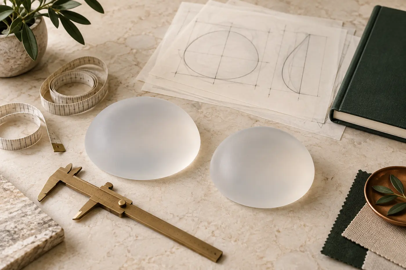
Breast base width, the distance between the breasts, sternum, rib-cage curvature and the existing inframammary fold help define safe pocket boundaries. If an implant base is poorly matched to the chest, changing the plane alone will not solve the mismatch. This is why implant volume, profile and base width must be planned with the pocket rather than after it.
Skin envelope, nipple position and ptosis
An implant adds volume; it does not reliably remove significant excess skin. Mild glandular descent and true breast ptosis are not the same. Nipple position in relation to the fold and the amount of loose skin influence whether augmentation alone or an approach involving a breast lift should be discussed. Dual plane can modify some tissue relationships but does not automatically replace a needed lift.
Muscle anatomy, occupation and sport
People who perform heavy resistance training, swim, climb or use pectoralis major intensively at work should discuss animation. This does not automatically rule out a subpectoral pocket or make subglandular placement correct. Visibility, coverage, performance priorities and expected movement need to be balanced.
Previous surgery and asymmetry
Existing incisions, an old implant pocket, capsule, tissue thinning and fold position can alter revision planning. The right and left sides may not begin alike; pocket details may occasionally need to differ. A breast asymmetry assessment should separate differences that may be reduced from those likely to remain.
Does thin tissue always require under-the-muscle placement?
No. Thin upper-pole coverage may make muscle coverage valuable, but implant size and profile, a subfascial option, supplemental fat grafting, skin quality and muscle use may also be considered. Fat grafting is not possible or sufficient for everyone; donor sites, variable fat retention and its own risks must be explained.
The reverse is also true: thicker tissue does not make an above-muscle pocket automatic. Breast shape, lower-pole anatomy, ptosis, desired volume and long-term tissue behaviour may change the recommendation. Sound planning does not use a “one measurement equals one method” formula.
What is animation deformity?
When pectoralis major contracts, an implant beneath it may move, flatten or shift outward. This is called animation deformity. Mild movement may only be visible during a particular exercise; in some people it is more noticeable in daily activity. Its possibility does not mean that every under-muscle augmentation fails, but it deserves specific discussion in people who train the chest heavily.
Severity depends on more than the label “submuscular”. Muscle release, pocket limits, implant dimensions and individual anatomy all contribute. For an existing implant with marked animation, conversion of the plane is not automatically the answer; capsule, tissue thickness, pocket and device condition must be examined.
Why can rippling and implant edges be more visible above the muscle?
Rippling means that folds in the implant shell can be seen or felt through the tissues. A thin envelope, an implant that is too large or poorly matched to the footprint, device characteristics, weight loss and pocket position can all contribute. Without muscle over the upper implant, this can be more apparent in some subglandular patients. Submuscular placement does not eliminate it, particularly along lower and outer portions not covered by muscle.
The solution is not simply a firmer or fuller implant. A better base–profile match, pocket correction, device exchange or selective fat grafting may be discussed. Each has limits and separate risks.
Does the plane alter capsular contracture risk?
The body forms a capsule around every implant. Capsular contracture occurs when this tissue tightens enough to make the breast firm, distort its shape or cause pain. Recent systematic reviews have reported higher rates with subglandular placement than with some submuscular approaches. However, studies differ in implant surfaces, surgical era, technique and length of follow-up, so one percentage cannot be applied to every patient.
Plane is only one variable. Infection or biofilm, haematoma, implant surface, incision, operative technique, previous radiotherapy and individual healing may also contribute. No pocket reduces the possibility of capsular contracture to zero.
What about displacement and double-bubble deformity?
An implant may shift down, up, inward or outward depending on pocket boundaries, tissue support, implant weight, fold anatomy and healing. A “double bubble” describes two visible lower contours formed by the old breast fold and the new implant contour. A constricted lower pole and the original fold position can affect this risk.
Dual-plane techniques may help manage particular lower-pole relationships but cannot make malposition impossible. Choosing a larger implant to “fill” loose skin can overload tissue if anatomical limits are exceeded.
Does the plane determine recovery time?
Techniques that elevate or stretch muscle may feel different in the early period, with pressure, tightness and discomfort during contraction. Muscle-related pain may be lower with an above-muscle pocket, but incision, implant size, a simultaneous lift, personal pain response and complications all affect recovery. The pocket alone cannot provide an exact return-to-work date.
Rapidly increasing one-sided swelling, uncontrolled pain, breathlessness, chest pain, fainting, fever, spreading redness or wound discharge should not be treated as routine recovery. Follow the surgical team’s written urgent-contact plan. Our first 30 days after breast surgery guide provides a general framework; your discharge instructions take priority.

Can dual plane replace a breast lift?
Not in every case. Implants add volume and alter some contours, but they do not remove substantial loose skin or reliably raise a low nipple in every anatomy. Mild gland descent must be distinguished from advanced skin excess. Our guide comparing breast lift and implants explains the difference between volume and position.
Combining augmentation with lift in one stage or separating the operations has distinct trade-offs. Trying to fill ptosis with excessive volume may stretch tissue further. Scars, blood supply, implant size and revision possibility require individual discussion.
Does implant placement change mammography and breast-health follow-up?
People with implants should continue routine or risk-based breast-health screening. Tell the imaging service about the implants before mammography so trained staff can use appropriate additional views. A new lump, spontaneous or bloody nipple discharge, skin tethering or unexplained one-sided change should not be postponed as an aesthetic concern.
Silicone implant rupture can be silent. The FDA describes MRI as the most effective way to detect silent rupture and ultrasound as an acceptable screening alternative for asymptomatic patients. Follow the device’s patient information and your clinician’s individual surveillance plan. Placement does not remove this long-term responsibility.
Are breast implants lifetime devices?
Breast implants should not be regarded as lifetime devices. Rupture, capsular contracture, malposition, rippling, pain, infection, tissue change or a change in preference may lead to further assessment and surgery. Equally, a fixed rule that every implant must be exchanged at ten years is not correct for everyone; symptoms, examination, imaging and device information guide the decision.
If an existing implant has become firm, changed shape, developed new asymmetry or caused pain, breast implant exchange or removal planning assesses the implant, capsule, pocket and natural tissue together. Knowing only the name of the old plane is insufficient.
Questions to ask your surgeon
- How did you assess my upper- and lower-pole tissue coverage?
- Where exactly will the pocket be, and which portions will have muscle coverage?
- Why does this plane fit my anatomy and priorities?
- What are my personal trade-offs for subglandular, submuscular, dual-plane or subfascial placement?
- How do my sport, work and pectoral activity affect animation?
- How will the plan address rippling, capsular contracture and malposition?
- Do I need a lift, and what can an implant alone not change?
- What device card, imaging, long-term review and revision plan will I receive?
Use our breast aesthetics consultation checklist to organise your health and implant records. An implant brand, volume or plane should not be fixed before a proper examination.
Can international patients choose a plane remotely?
A video consultation can collect history and goals, but tissue thickness, inframammary fold, chest wall, muscle movement and the degree of ptosis need in-person examination. Before travelling to Antalya, understand that a preliminary conversation does not confirm an operation or device decision.
Allow flexible time for assessment, any imaging or tests, surgery, early reviews and safe return. The technique should not be forced into a travel timetable; travel should be organised around a safe clinical process.
Op. Dr. Bahadır Çelik’s planning approach
During a breast augmentation assessment with Op. Dr. Bahadır Çelik, the implant plane is not selected in isolation. Chest wall, breast footprint, upper- and lower-pole tissue, nipple position, asymmetry, muscle activity, implant dimensions and long-term follow-up belong to one plan. The aim is not to apply one named technique to everyone, but to explain the individual rationale, alternatives and limits.

Conclusion: the right plane needs the right reason
Subglandular, submuscular and dual plane are not a ranking from bad to good. An above-muscle pocket may be less affected by contraction but requires adequate coverage. A subpectoral pocket can add upper coverage but introduces muscle interaction. Dual plane aims to balance upper coverage and lower-pole tissue behaviour, yet it is neither universal nor complication-free.
The decision begins by defining anatomy and priorities before implant volume. A useful consultation should leave you able to explain where the proposed pocket is, why it was recommended, the alternatives, what it cannot change and the long-term responsibilities in your own words.
Medical note: This article is general information, not a personal diagnosis or surgical recommendation. Seek timely medical assessment for a new breast lump, bloody discharge, skin change, persistent swelling, fever, rapid one-sided enlargement or late fluid around an implant.
References
- American Society of Plastic Surgeons — Breast augmentation procedure and implant placement
- International Society of Aesthetic Plastic Surgery — Breast augmentation
- BAPRAS — Breast implant techniques
- US Food and Drug Administration — Breast implants
- FDA — Risks and complications of breast implants
- Tebbetts JB — Dual plane breast augmentation and implant–soft-tissue relationships
- Alderhali et al. — Implant pocket plane selection in primary breast augmentation: systematic review and network meta-analysis
- Haas et al. — Capsular contracture after breast augmentation: systematic review and meta-analysis
- Yuan et al. — Outcomes in subfascial versus subglandular breast augmentation: systematic review
- Republic of Türkiye Ministry of Health — Regulation on healthcare promotion and information
FAQ
Frequently asked questions
Is a breast implant better under or over the muscle?+
What exactly does dual-plane breast augmentation mean?+
Must very slim patients have implants under the muscle?+
Is recovery faster with over-the-muscle implants?+
What is animation deformity with under-muscle implants?+
Does subglandular placement increase capsular contracture risk?+
Can dual plane replace a breast lift?+
Does implant position change mammography?+
Related information
Implant Planning Guide · 16 min read
Why Volume Alone Is Not Enough When Choosing Breast Implants
Read article↗
Postoperative Recovery Guide · 17 min read
The First 30 Days After Breast Surgery: A Recovery Guide
Read article↗
Lift and Volume Decision Guide · 19 min read