Male Breast Surgery · Antalya
Gynecomastia Surgery
A detailed guide to gland versus fat, investigation of causes, liposuction and excision, scars, risks, recovery and structured follow-up.
Results of every surgical or invasive procedure may vary from person to person. You are advised to obtain detailed information from your doctor before the procedure.
The cover and planning images on this page are illustrative; they do not show a real patient or a treatment result.
Gynecomastia surgery is not one standard operation for every enlargement of the male chest. Gland behind the nipple, surrounding fat, skin excess, the pectoral muscle and the rib cage must be assessed separately. If enlargement is new, painful or associated with a firm one-sided lump, nipple discharge or signs of a hormonal disorder, the cause should be investigated before aesthetic planning. Surgery is considered only after the diagnostic findings, individual goals and tissue characteristics have been reviewed together.
In brief: Gynecomastia means benign enlargement of glandular breast tissue in a male; fat accumulation alone is not the same condition. Weight loss may reduce the fatty component but does not reliably remove established gland. Management can include observation, treatment of an underlying cause, selected medical therapy for recent cases and, when appropriate, liposuction, gland excision or a combined operation.
What is gynecomastia?
Gynecomastia is benign growth of glandular tissue beneath the nipple and areola in a male chest. It may affect one or both sides, and two-sided enlargement does not have to be equal. Tenderness, fullness or a rubbery disc beneath the nipple may be noticed. Fat and loose skin can accompany the gland and alter the visible contour.
The word describes a clinical finding rather than every appearance of a prominent male breast. Enlargement caused mainly by fat, gland, loose skin, a distinct mass or a combination of these findings leads to different investigations and treatment choices. A procedure cannot therefore be selected from an internet photograph or body measurement alone.
How do true gynecomastia, pseudogynecomastia and loose skin differ?
True gynecomastia contains palpable glandular tissue, usually centred beneath the nipple. In pseudogynecomastia, also called lipomastia, fat is the main component and may become more apparent with weight gain. Long-standing volume, major weight loss or ageing may add loose skin and a lower nipple position.
The distinction is more detailed than deciding whether the chest feels hard or soft. Gland may be localised or broad; fat can extend towards the sides of the chest; and skin elasticity may be strong or limited. Pectoral muscle volume and the shape of the ribs also influence symmetry. Planning is based on which layer creates the contour and how much each layer contributes.
What causes gynecomastia?
A shift in the balance between oestrogenic and androgenic effects on breast tissue can promote gland growth. Gynecomastia occurs during normal physiological periods such as infancy, puberty and later life. Even when there appears to be an obvious reason for new adult-onset enlargement, a careful history and examination remain important.
- Temporary hormonal changes during puberty
- Low testosterone or conditions affecting testicular function
- Thyroid, liver, kidney and other systemic disorders
- Rare hormone-producing tumours of the testis or another organ
- Certain prescription medicines, hormones, herbal products and sports supplements
- Anabolic-androgenic steroids and some recreational substances
- Marked weight gain, residual skin after weight loss or no identifiable cause
A medicine appearing on a list associated with gynecomastia is not a reason to stop it without advice. Changes to prescribed treatment should be coordinated with the clinician responsible for it. Likewise, one “normal hormone test” does not automatically exclude every cause; the choice and interpretation of tests depend on the clinical findings.
How is adolescent gynecomastia approached?
Pubertal gynecomastia is common and often improves over time. Recent, typical enlargement without warning signs can usually be observed while growth and puberty progress. Rapid growth, persistence, substantial pain or serious psychosocial distress nevertheless deserves medical assessment rather than dismissal.
A decision about adolescent surgery is not based on age alone. Duration and stability of enlargement, investigation of a cause, weight change, substance use, physical symptoms, maturity for informed decision-making and the possibility of recurrence are considered. Parental permission is important for a minor but does not replace the young person’s views or an assessment of medical appropriateness.
Which symptoms should not wait for a routine cosmetic consultation?
A new or rapidly enlarging firm one-sided mass, especially bloody nipple discharge, nipple inversion, skin dimpling or ulceration, an underarm lump, pronounced redness, unexplained weight loss or a testicular mass requires prompt medical assessment. Male breast cancer is uncommon, but a male breast lump should not automatically be labelled gynecomastia.
New severe pain, fever, rapidly increasing swelling after trauma or signs of infection also fall outside routine aesthetic planning. These symptoms do not automatically mean cancer; infection, a haematoma, a cyst and other benign conditions are possible. The diagnosis comes first.
What information is discussed at the first consultation?
The history includes when the enlargement began, whether one or both sides are involved, the rate of change, pain or tenderness, nipple discharge, weight history and pubertal development. Sexual function, libido, fertility plans, testicular disease or surgery, and a history of thyroid, liver or kidney disease may guide the medical assessment.
A complete list of prescription and non-prescription medicines, hair-loss or prostate products, hormones, sports supplements and herbal products is important. Anabolic steroids and other substances should be discussed openly and without judgement. Previous laboratory and imaging reports can be brought to the appointment. Prescribed medicines should not be paused without the relevant clinician’s advice.
What is assessed during physical examination?
The examination considers whether gland is present, its diameter, consistency and relationship to the nipple, together with fat distribution, loose skin, areolar diameter and nipple level. The clinician assesses whether a mass is central or separate from the nipple and irregular, and looks for associated skin or underarm findings. Pectoral muscle and chest-wall asymmetry are also documented.
When indicated, examination may include the testes and signs of thyroid or liver disease or androgen deficiency. Standardised photographs, measurements and body-mass index can support planning, but none replaces an examination and appropriate diagnostic assessment.
Does every patient need hormone and blood tests?
Testing is individualised according to the history and examination. Unexplained new adult-onset enlargement or relevant symptoms may lead to assessment of testosterone, estradiol, LH, FSH, thyroid function, hCG and liver or kidney function. Ordering the same panel for everyone, or making a surgical decision from a single result, is not appropriate.
An abnormal result may require evaluation by endocrinology, urology or another specialty. Treating an underlying disease or medication effect concerns general health as well as the chest appearance. Removing tissue surgically does not correct an untreated hormonal or systemic cause.
When are ultrasound, mammography or biopsy considered?
Routine imaging is generally not recommended when examination is typical of gynecomastia or pseudogynecomastia. If a mass is indeterminate or the examination is suspicious for malignancy, ultrasound, mammography or digital breast tomosynthesis may be selected according to age and findings. The ACR approach commonly starts with ultrasound for an indeterminate mass in a man younger than 25 and mammography or tomosynthesis at 25 or older; suspicious examination findings can justify mammography or tomosynthesis at any age.
Imaging is used to clarify the nature of a mass, not to produce a surgical marketing image. If the findings remain indeterminate or suspicious, needle biopsy or tissue sampling may be necessary. A cosmetic procedure should not precede completion of this diagnostic pathway.

How are the treatment pathways separated?
| Approach | When might it be considered? | Important limitation |
|---|---|---|
| Observation | Typical pubertal gynecomastia or a recent case that may regress | Warning signs and an underlying disease must first be considered |
| Cause-directed care | A medicine, hormonal issue or systemic disease is identified | Prescription treatment is not stopped independently; established gland may persist |
| Selected medical therapy | Some recent, painful cases judged suitable by the relevant specialist | Long-standing fibrotic tissue may respond poorly; self-medication is unsafe |
| Liposuction | Fat predominates and skin is expected to retract | It may not adequately remove firm gland |
| Gland excision | A distinct firm gland is present behind the nipple | Over-resection can cause a depression; under-resection can leave fullness |
| Combined surgery and skin adjustment | Gland, broad fat and/or substantial loose skin occur together | Additional scars, recovery and blood-supply risks may be introduced |
Can exercise and weight loss remove gynecomastia?
Weight loss and regular exercise may reduce overall body fat and the fatty component of the chest. Developing the pectoral muscle changes proportions, but exercise does not selectively dissolve glandular breast tissue. A larger muscle can occasionally make the overlying gland more visible.
For someone above a sustainable weight, weight management can reveal the true surgical need and probable skin behaviour more accurately. However, telling every person simply to reach an “ideal weight” is not a complete assessment. Gland, pain, medication history and warning signs should be assessed independently of weight.
Is non-surgical or medical treatment possible?
When a cause is identified, treating the condition or safely changing an associated medicine is the first step. In selected recent and tender cases, medical treatment may be considered by endocrinology or another appropriate specialist. Using anti-oestrogen or hormone-modifying drugs obtained online can affect clotting, the liver, hormones and reproductive health.
Long-standing gland tends to become more fibrotic and less likely to shrink with medicine. Fat-freezing, energy devices and creams do not reliably eliminate true gland. When a non-surgical option is discussed, the type of tissue and the limits of the evidence should be made clear.
Who may be considered for gynecomastia surgery?
A surgical consultation may be appropriate when the cause has been assessed, chest development has remained stable, weight is reasonably steady, nicotine and substance use can be managed safely, and the person understands scars, risks and alternatives. Physical discomfort, difficulty with clothing or exercise, and persistent psychosocial burden may all contribute to the decision; social pressure alone does not make surgery necessary.
Surgery may be postponed if there is uncontrolled systemic disease, active infection, incomplete diagnostic assessment, ongoing anabolic-steroid use, major weight fluctuation or unrealistic expectations. The objective is not perfect symmetry or creation of a muscular chest. It is a proportionate reduction of excess gland and fat within the limits of the individual anatomy.
When is liposuction used?
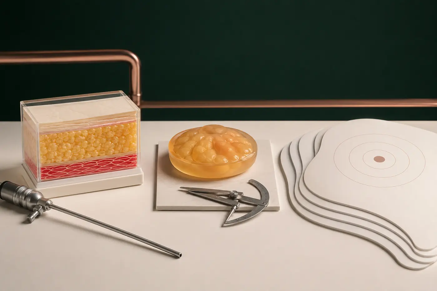
Liposuction reduces fat through small access points using a cannula. The front of the chest, lateral extension and transition into neighbouring areas are considered together. The exact technique varies with skin thickness, the fibrous nature of the fat and the surgical plan.
Conventional liposuction does not reliably remove firm gland. Treating fat alone can leave fullness beneath the nipple, while overly superficial or irregular treatment can produce rippling, adherence or depressions. Liposuction may be used alone for a predominantly fatty chest or combined with direct gland excision.
How is glandular tissue removed?
A distinct gland is commonly removed directly through a limited incision at the edge of the areola. Incision position and length depend on gland size, distribution and any additional procedure. An appropriate layer is preserved beneath the nipple–areola to support the contour, while the edges are blended with the surrounding chest tissue.
Removing “100 per cent of the gland” is not the correct goal in every case. Excessive removal may cause a crater deformity, adherence or blood-supply problems. Inadequate or uneven removal may leave residual fullness and asymmetry. The amount is adjusted to tissue thickness, contour and circulation during the operation.
Combined treatment, skin removal and nipple position
When gland and fat coexist, liposuction and direct excision can be combined. Skin may gradually adapt after a substantial volume reduction if elasticity is good. Marked skin excess, a large areola or a low nipple may instead require skin removal and areolar adjustment.
Skin incisions may remain around the areola or extend horizontally and vertically in more advanced cases. A staged operation is sometimes considered to protect the blood supply. The balance between leaving extra skin to keep scars shorter and accepting longer scars to improve the contour should be discussed explicitly.
How are anaesthesia and the surgical setting planned?
Depending on the extent of treatment and the patient’s health, general anaesthesia or, for selected limited procedures, sedation with local anaesthesia may be discussed. Anaesthesia is not selected by the patient alone; the surgeon and anaesthetist determine a safe plan. Surgery is performed in a licensed healthcare facility under sterile conditions.
Operating time varies between liposuction alone, combined gland excision and extensive skin adjustment. A drain is not mandatory in every case. It may be used temporarily according to the dead space, risk of bleeding or fluid accumulation and the surgeon’s technique. Same-day discharge or overnight observation depends on the clinical situation.
Where are the scars, and is tissue sent to pathology?
Liposuction access points are small but still leave scars. For gland excision, the scar is often placed along the colour transition of the areola; skin removal requires longer scars. Scars are permanent and can initially look pink, dark or firm. Genetics, nicotine, infection, tension and aftercare influence maturation.
Whether excised gland is sent for histopathology depends on age, examination and imaging findings, the appearance of the tissue and local clinical policy. Histology becomes particularly important with an atypical or suspicious finding. It should not be used to delay a necessary pre-operative biopsy.
How should a person prepare for surgery?
- Disclose every medicine, hormone, supplement and recreational substance.
- Complete the diagnostic pathway if there is a suspicious mass or hormonal sign.
- Adjust prescription medicines only under the relevant clinician’s plan.
- Agree an individual timetable for stopping smoking, vaping and nicotine products.
- Discuss stabilisation if body weight is still changing.
- Share bleeding, clotting and anaesthetic history, allergies and previous operations.
- Clarify scars, a possible drain, compression garment and home support in advance.
Pre-operative markings may be made while standing so existing differences between the two sides are recorded. Goals such as “completely flat,” “scarless” or “exactly identical on both sides” are not medically realistic. Consent should include the alternatives and the option of having no procedure.
What happens during recovery?
| Period | Changes that may occur | General approach |
|---|---|---|
| First 72 hours | Swelling, bruising, tightness, soreness and unequal appearance | Short walks, prescribed medicines and monitoring for bleeding or circulation problems |
| First 1–2 weeks | Oedema, temporary firmness, altered sensation and possible drain care | Wound care, scheduled review, advised compression and no heavy lifting |
| Weeks 2–6 | The contour becomes clearer; firmness or rippling can persist | Gradual return to work and exercise according to individual instructions |
| Following months | Swelling settles, tissue softens and scars mature | Do not judge the final result early; continue required reviews |
ASPS patient information notes that the final contour may take three to six months, and extensive skin procedures or individual healing can alter that timeline. Early firmness, tethering or asymmetry beneath the nipple may change as swelling settles. Massage, devices or medicines should not be started without examination and advice.

How long is compression worn, and when can work and sport resume?
A compression garment may support swelling and protect the area under clothing. Its pressure and duration vary with the procedure. A garment that is too tight can cause circulation and skin problems. The surgical team explains when it may be removed, washed and whether it is used at night.
Desk work, heavy manual work, swimming, chest training and contact sport do not restart at the same time. Driving requires discontinuation of sedating pain medicine, safe arm movement and the ability to brake suddenly. Flight and long-distance travel plans should account for clot risk, the extent of surgery and the review schedule.
Which postoperative symptoms require urgent assessment?
Rapidly increasing swelling and tension on one side, bleeding that soaks the dressing, worsening pain, a dark or very pale nipple, high fever, spreading redness, foul discharge, shortness of breath, chest pain or swelling in one leg can require urgent assessment. If a drain is present, sudden cessation, a sharp increase in bright-red fluid or accidental displacement should also be reported.
What are the risks of gynecomastia surgery?
General risks include anaesthetic complications, bleeding and haematoma, infection, seroma, blood clots, poor wound healing, conspicuous scars, persistent pain and the possibility of further surgery. Nipple sensation may increase or decrease temporarily or permanently. Areolar asymmetry, fat necrosis, impaired skin or nipple circulation and, rarely, tissue loss may occur.
Procedure-specific concerns include contour irregularity, depressions, tethering, rippling, different volume on each side, nipple inversion, persistent skin excess or residual gland. Liposuction, gland excision and skin removal each have a different risk profile. A possible need for revision cannot always be judged in the early healing phase.
What causes a crater deformity, residual fullness or recurrence?
Removing too much tissue beneath the nipple can create a crater-like depression and adherence to the underlying muscle. Inadequate or uneven excision can leave fullness. Swelling must be allowed to settle before residual tissue is distinguished from temporary postoperative change, and revision should not be planned without examination at an appropriate time.
Excised gland does not simply grow back in full, but remaining tissue can become prominent again with hormonal changes, weight gain, continued anabolic-steroid exposure or an associated medicine. Surgery does not provide lifelong protection from future weight or endocrine changes.
What about sensation, sexual function, fertility and future breast health?
Gynecomastia surgery does not operate on the testes or sperm ducts. However, an underlying hormonal cause can relate to sexual function and fertility. Reduced libido, erectile problems, infertility, small testes or plans for children should therefore be included in the history. Hormone medicines should not be used without specialist assessment.
Nipple sensation can change temporarily or permanently after surgery. Gynecomastia is not itself breast cancer, and most male breast changes are benign. Nevertheless, a future new lump, discharge, skin change or nipple change should not be ignored simply because surgery has already been performed.
Consultation in Antalya and follow-up after travel
For a gynecomastia consultation in Antalya, a person can prepare a list of medicines and supplements, available hormone results, ultrasound or mammography reports and details of previous operations. An online appointment may help with history-taking and travel planning, but it cannot replace examination of the gland, skin, a mass or the testes. The final surgical plan follows an in-person assessment.
For patients travelling to Antalya, the licensed operating facility, completion of investigations, anaesthetic assessment, local stay, drain and review schedule, flights or long journeys, urgent contact and follow-up after returning home should be documented. Travel is arranged around recovery; surgery should not be planned as a holiday activity.
Useful questions to ask during consultation
- How much of the contour comes from gland, fat, skin or chest-wall difference?
- Do I need blood tests, testicular assessment or another specialist opinion?
- Is the examination typical, or are ultrasound, mammography or biopsy indicated?
- Why would I need liposuction, gland excision or skin adjustment?
- Where and how long will the scars be, and will tissue be sent to pathology?
- How will my risks of a crater, residual fullness, asymmetry and nipple blood-supply problems be managed?
- What are my individual schedules for compression, drains, work, driving, sport, swimming and travel?
- Which symptoms are urgent, and how can I reach the team?
Medical responsibility for this content

This page is prepared for general information using current andrology, male-breast imaging and plastic-surgery sources and is medically reviewed by Op. Dr. Bahadır Çelik before publication. The review date shown on the page records the completed clinician review. The content does not replace examination, diagnosis, informed consent or an individual treatment plan.
References
- European Academy of Andrology — Clinical Practice Guidelines on Gynaecomastia
- American College of Radiology — Evaluation of the Symptomatic Male Breast
- Endotext — Gynecomastia: Etiology, Diagnosis and Treatment
- National Cancer Institute — Breast Cancer Signs and Symptoms
- American Society of Plastic Surgeons — Gynecomastia Surgery Candidates
- American Society of Plastic Surgeons — Gynecomastia Surgery Procedure
- American Society of Plastic Surgeons — Gynecomastia Surgery Safety and Risks
- American Society of Plastic Surgeons — Gynecomastia Surgery Recovery
- American Society of Plastic Surgeons — Gynecomastia Surgery Results
- Republic of Türkiye Ministry of Health — Regulation on Health-Service Promotion and Information
FAQ
Frequently asked questions
Can exercise or weight loss completely remove gynecomastia?+
Does one-sided gynecomastia mean cancer?+
Does everyone need hormone tests and imaging before gynecomastia surgery?+
Is liposuction alone enough for gynecomastia?+
Does gynecomastia surgery leave scars?+
How long is compression worn, and when can exercise restart?+
Can gynecomastia return after surgery?+
When may gynecomastia surgery be considered for an adolescent?+
Related information
 01
01Breast Augmentation
A comprehensive guide to implants, fat transfer and, where appropriate, breast lift planning based on chest anatomy, tissue coverage, asymmetry, expectations and long-term follow-up.
Read article↗ 01
01Breast Lift Surgery
A comprehensive guide to nipple position, excess skin, existing tissue, scar patterns, implant decisions, recovery, risks and long-term change after a breast lift.
Read article↗ 01
01Breast Reduction Surgery
A detailed guide to symptoms associated with heavy breasts, tissue and incision options, scars, breastfeeding, recovery, risks and long-term follow-up.
Read article↗