Gland and Fat Assessment Guide
Gynecomastia or Chest Fat? How the Difference Is Assessed
Understand glandular gynecomastia versus chest fat, evaluation of causes and red flags, and when liposuction or direct gland excision may have a role.
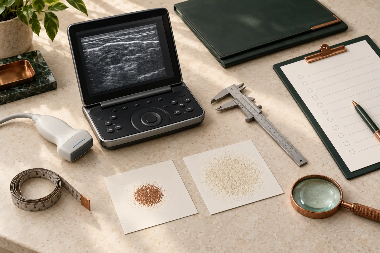
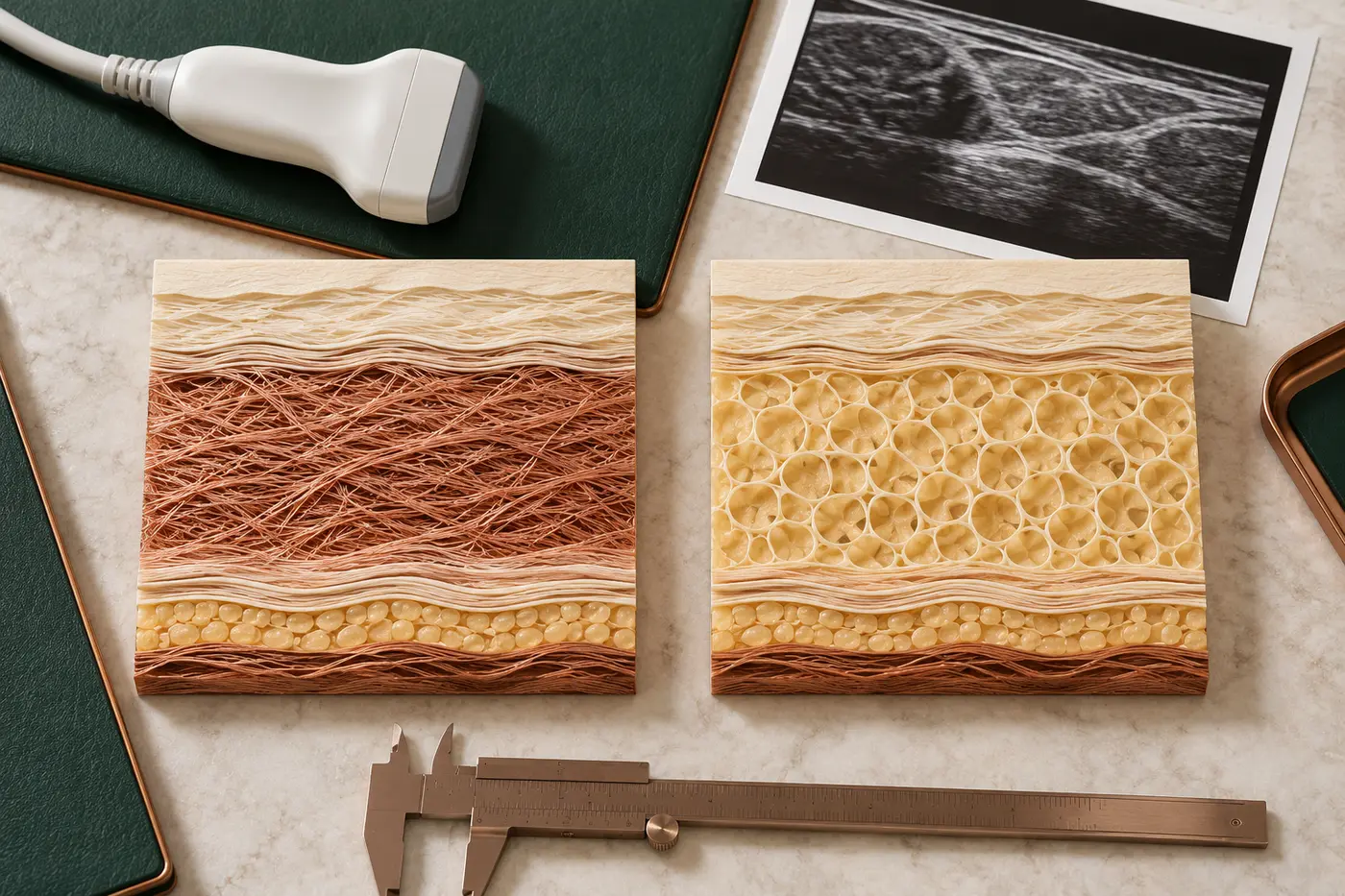
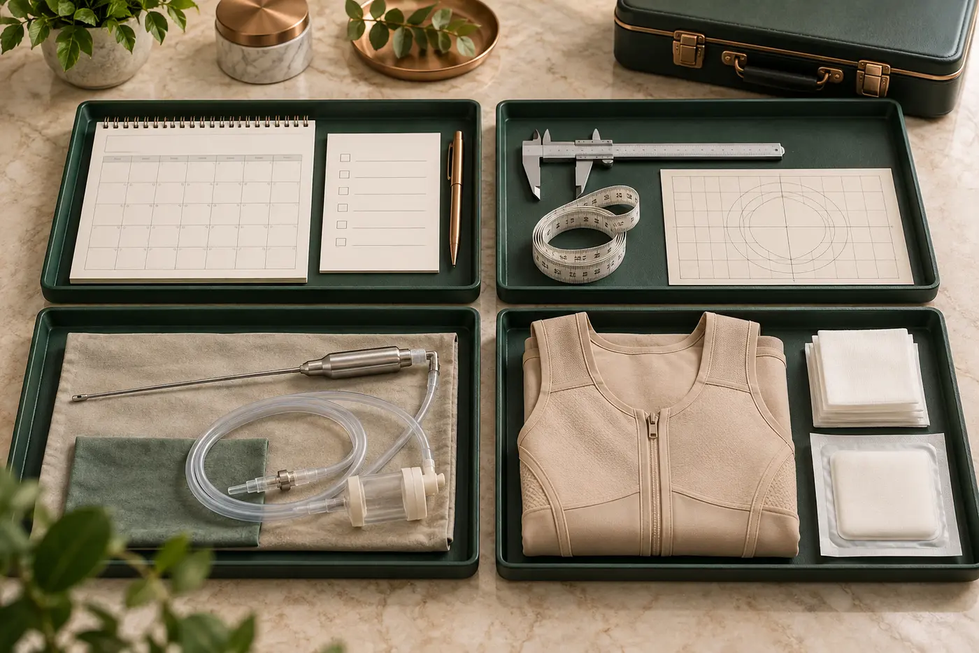
Image note: The clinical editorial visuals on this page show no patient, real body, operation or treatment result. Abstract models explain the gland-versus-fat distinction and the assessment pathway.
Short answer: Gynecomastia is benign growth of glandular breast tissue in a male, usually centred behind the nipple. Chest fat is predominantly an increase in adipose tissue and may be called pseudogynecomastia or lipomastia. Both components can occur together. A photograph, a home “pinch test” or body weight alone cannot distinguish them reliably. History and examination come first; targeted laboratory tests or imaging are added when the findings are unclear or concerning.
Gynecomastia or chest fat: what is the difference?
Fullness of the male chest does not have a single cause. In true gynecomastia, glandular tissue proliferates behind the nipple–areola complex. In pseudogynecomastia, adipose tissue accounts for most of the contour. A common third pattern is mixed: a central glandular component sits within a wider layer of fat.
This distinction is more than terminology. It affects how much weight management may change the contour, whether an underlying cause needs investigation, and the relative roles of liposuction and direct gland excision. Even when gland is felt, however, this is not automatically an indication for surgery. Onset, duration, medicines, puberty, weight, hormonal features and systemic symptoms also matter.
Why is appearance alone unreliable?
A mirror or photograph cannot measure the proportion of gland and fat. Diffuse fat under tight skin may feel surprisingly firm, while longstanding gland blends with surrounding adipose tissue. Cold, muscle tension and camera angle can temporarily change areolar projection. A developed pectoral muscle also alters the chest silhouette.
The online “two-finger pinch test” is not a diagnostic method. Forceful repeated pinching can cause pain and cannot safely exclude a malignant lump. A one-sided, hard, irregular, rapidly enlarging or off-centre mass, or a change in the skin or nipple, requires a medical diagnostic pathway before aesthetic planning.
How do glandular and fatty tissue feel?
Glandular tissue is often described as a denser, rubbery disc or button immediately behind the areola. Fat tends to be softer and more diffuse, sometimes extending towards the side of the chest and underarm. Palpation varies between individuals and cannot provide a perfect percentage of each tissue.
The examination may be performed standing and lying down. Nipple position, areolar diameter, excess skin, pectoral muscle, side-to-side difference and the axilla are assessed. The useful question is not only “is gland present?” but “which layers create this contour?”
What causes gynecomastia?
Gynecomastia may develop when the balance of oestrogenic and androgenic activity at breast tissue changes. This does not always mean that a blood oestrogen value is high or testosterone is low. Tissue sensitivity, hormone production and conversion, medicines or substances and the stage of life can all contribute.
Physiological stages of life
Gynecomastia is more common in newborns, during puberty and in older age. Most pubertal cases regress over time, so a recent, clinically reassuring presentation in an adolescent does not automatically call for surgery. Duration, tenderness, growth and psychosocial effect are followed.
Weight and body composition
Higher body fat can enlarge the chest and make an existing glandular component more visible. Hormone conversion within adipose tissue may also contribute to the biological context. Yet not every enlarged chest in an overweight person is fat alone, and a lean person can have both gland and local fat.
Medicines and substances
Some hormonal and antiandrogen medicines, selected psychiatric and cardiovascular drugs, anabolic steroids and other substances have been associated with gynecomastia. An association does not prove that a particular drug caused the finding. Prescribed treatment must not be stopped without the prescribing clinician, and supplements, sports products and substance use should be disclosed without fear of judgement.
Endocrine and systemic conditions
Testicular, thyroid, liver or kidney disease, proven testosterone deficiency and rare hormone-producing tumours may enter the differential diagnosis. An underlying disorder should be considered in adult-onset gynecomastia, but ordering the same broad hormone panel for everyone is not good assessment. Tests are selected from the history and examination.
Which findings should not be delayed?
Male breast cancer is uncommon, and gynecomastia is not considered a premalignant condition. Nevertheless, the following findings should not be labelled “just gynecomastia” without assessment:
- A one-sided, hard, irregular or off-centre breast lump,
- Nipple retraction, bloody or clear discharge, or a non-healing sore,
- Skin dimpling, orange-peel change, persistent redness or ulceration,
- A hard underarm swelling,
- Rapid growth, unexplained weight loss or systemic illness,
- A testicular lump, marked testicular change, pain or a new change in sexual function.
These signs do not themselves diagnose cancer. They indicate that the correct medical assessment should come before cosmetic surgery. Suspicious findings may require tailored imaging and direct core biopsy rather than repeated reassurance or blind excision.
What is asked in the medical history?
The clinician records age at onset, duration, growth rate, pain or tenderness, one- or two-sided involvement, weight change and family history. Pubertal development, libido and sexual function, fertility history, testicular symptoms and signs of systemic disease are discussed with privacy and a clear explanation.
Prescribed and over-the-counter medicines, testosterone or anabolic steroids, herbal products, sports supplements, alcohol and other substances can be relevant. Finding an apparent cause does not make the rest of the examination unnecessary; a second issue may still need to be excluded.
Use the consultation preparation checklist to organise product packaging, previous tests and a timeline of the change. Shared photographs do not replace medical history or face-to-face examination.

How are examination and laboratory tests planned?
Breast examination aims to confirm a glandular component, recognise diffuse fat, identify a suspicious mass and inspect the skin and nipples. When indicated, the genital examination and signs of thyroid, liver or other systemic disease are also assessed. Privacy, explanation and consent are essential throughout.
Depending on the findings, selected tests may include testosterone, oestradiol, LH, FSH, SHBG, TSH, prolactin, hCG, AFP and liver or renal function. The European Academy of Andrology guideline lists these as a possible evaluation set; it does not mean that every person requires every test. Collection time, fasting state, medicines and the laboratory method can affect some results.
Does everyone need ultrasound or mammography?
No. Imaging is not automatically required when the clinical examination is typical and reassuring. Ultrasound or age- and finding-appropriate breast imaging may be used when the examination is equivocal, a lump is one-sided or suspicious, or anatomy needs clarification before surgery. A suspicious lesion should be managed with breast radiology and, when indicated, a core biopsy.
Ultrasound is not a perfect aesthetic map that produces a precise “gland percentage”. It must be interpreted alongside the examination. A normal ultrasound also does not automatically establish surgical suitability or select a technique.
Will weight loss reduce the chest?
If fat is a major component, sustainable weight loss can reduce chest circumference and reveal the underlying contour. Established glandular tissue cannot be selectively burned away by exercise. Chest training develops the pectoral muscle; the gland and fat above it behave differently. In some physiques, a larger muscle may make overlying fullness appear more pronounced.
When weight is changing rapidly, surgical measurements may not represent the stable situation. Moving towards a realistic, maintainable weight helps assess anaesthetic risk and potential skin excess. Gynecomastia surgery is not an obesity treatment or a general weight-loss method.
Why is the approach different in adolescents?
Pubertal gynecomastia is common, and the great majority of cases may regress within 24 months. Observation is therefore usually the first step for a recent, stable and clinically reassuring presentation. Pain, rapid growth, marked asymmetry, substance use and psychosocial effect still deserve attention.
When the condition persists and causes substantial distress, paediatric/adolescent health, endocrinology and surgical assessment may need to work together. The decision should consider development, the young person’s capacity for consent, realistic expectations and body image rather than being driven solely by a parent or by social pressure.
What are the non-surgical options?
Observation may follow treatment of an underlying disease or supervised discontinuation of an associated substance. In overweight individuals, lifestyle and weight management may reduce the fatty component. Selected early and painful cases may be considered for medical treatment by the relevant specialist; self-prescribing hormones or “oestrogen blockers” bought online is unsafe.
Testosterone is not a treatment unless deficiency has been properly demonstrated and managed. Unsupervised testosterone or anabolic steroid use can worsen the problem. Longstanding fibrotic glandular tissue may respond poorly to medication. A compression garment can disguise the contour under clothing but does not remove tissue.
When may gynecomastia surgery be considered?
After reversible and underlying causes have been assessed, surgery may be considered for longstanding gynecomastia that does not regress and causes physical or meaningful psychosocial discomfort. The goal is not simply to remove as much tissue as possible; it is to create a contour that fits the chest while avoiding tethering, craters and irregularity.
Planning for gynecomastia surgery separately assesses fat distribution, glandular tissue, skin excess, the areola, nipple position and the pectoral framework. There is no single standard technique for every chest.
Liposuction alone
When fullness is mainly fatty and skin elasticity is favourable, liposuction may reduce the contour. A firm central gland may not be removed sufficiently with a cannula. Liposuction is not scarless; it uses small access incisions. Aggressive or superficial fat removal can create depressions and waviness.
Direct gland excision
A prominent glandular component often needs direct excision through a limited incision at the areolar border. Leaving a controlled thickness beneath the nipple helps reduce adherence and a crater deformity. Leaving too much can preserve a bulge, so the balance is made from preoperative assessment and intraoperative contour.
Combined liposuction and excision
For a mixed pattern, a common approach is to blend the surrounding fat with liposuction and remove the central dense gland directly. These are not competing techniques; they address different tissue layers. The distribution of tissue, not a branded operation name, should define the plan.
Managing excess skin
After a large volume, advanced descent or major weight loss, the skin may not contract enough. Skin excision and nipple repositioning through periareolar or longer scars can be necessary. A wish for the shortest scar should not override the tissue management required for a safe and even contour.
How do the options compare?
| Approach | Main component addressed | Key limitation |
|---|---|---|
| Observation / treating a cause | Recent, physiological or potentially reversible presentation | Longstanding fibrotic gland may not fully regress |
| Weight management | Diffuse fat and general health | Does not selectively remove glandular tissue |
| Liposuction | Fat-dominant contour | May be insufficient for dense central gland |
| Direct gland excision | Dense tissue behind the areola | Scar, bleeding, crater and altered nipple sensation risks |
| Combined technique | Mixed glandular and fatty pattern | Requires balance across two tissue layers and a broader recovery |
| Skin excision | Substantial laxity and descent | Longer and more visible scars |
This table cannot select an operation for an individual. The same apparent volume may need a different plan because skin, muscle, gland and fat distribution differ.
Where are the scars?
Liposuction uses access points that leave small scars. Gland excision is commonly placed along the lower areolar border. Excess-skin surgery may require a scar around the areola and longer horizontal or vertical scars. Colour and thickening vary with genetics, skin type, tension, infection, sun and nicotine.
“Scar-free gynecomastia surgery” is not a reliable claim. The aim is to use appropriately placed scars for the required correction. Scar care is individual, and no cream can guarantee complete disappearance.
What are the risks?
Bleeding and haematoma, infection, fluid collection, delayed healing, wide or raised scars, asymmetry, residual fullness, over-resection and a crater, contour waviness, altered nipple sensation or circulation, pain and revision may occur. A haematoma can present as rapidly increasing one-sided swelling and pain soon after surgery and needs urgent assessment.
Nicotine, poorly controlled health conditions, medicines or supplements affecting bleeding and anabolic steroid use can change risk. Do not stop prescribed medicines without a plan. Honest disclosure of substances supports safer anaesthesia and assessment of recurrence.
How does recovery progress?
Swelling, bruising, tightness and temporary asymmetry are expected initially. The duration of compression, dressings, showering, arm movement, driving and return to work or training depends on the procedure. Liposuction alone does not have the same recovery burden as gland excision plus skin removal.
Swelling falls over weeks, while skin adaptation and scar maturation take months. An early photograph should not be used to judge the final contour. Rapidly increasing one-sided swelling, uncontrolled pain, breathlessness, fever, spreading redness or wound discharge requires immediate contact.
For a general timeline and warning signs, see the first 30 days after breast surgery guide; your individual discharge instructions always take priority.
Can gynecomastia return?
Marked recurrence is not common after appropriate tissue reduction, but weight gain, anabolic steroids, associated medicines or substances and an untreated endocrine or systemic cause can change the contour again. Surgery cannot remove every gland cell or eliminate lifetime biological change.
Completing the causal assessment before surgery and maintaining a sustainable weight and medicine/substance plan afterwards are important. Early residual swelling should not be mistaken for recurrence; tissues need time to settle before formal review.
What should international patients plan in Antalya?
Remote photographs can show an overall contour but cannot confirm the gland–fat distinction, exclude a lump or measure skin quality. If there is a diagnostic question, allow time before surgery for the appropriate examination, laboratory work and imaging. Flights and hotels should not be made inflexible before the assessment is complete.
Early review, the risk of haematoma, compression and return travel need a joined plan. Ask for individual timing for heavy luggage, upper-body training, swimming and sun. Access to local healthcare after returning home and the method of contacting the clinic should be clear.
Questions to ask at the consultation
- How much of my contour comes from gland, fat, muscle or excess skin?
- Is there a one-sided or suspicious finding that needs imaging first?
- Do my medicines, supplements, steroid use or health history need further assessment?
- Should I first observe the condition or stabilise my weight?
- Why would liposuction alone be sufficient or insufficient?
- How much tissue is left beneath the nipple, and how is a crater avoided?
- Will skin excision be needed, and where will the scars be?
- Which early postoperative findings require urgent contact?
Op. Dr. Bahadır Çelik’s assessment approach
In an assessment with Op. Dr. Bahadır Çelik, “gynecomastia or fat?” is not reduced to a single label. Gland, fat, muscle, skin, areola and the chest wall are considered separately. A new or suspicious clinical finding is directed to the appropriate diagnostic pathway before any aesthetic operation.
Once reversible causes and weight change have been clarified, the roles of observation, liposuction, gland excision and skin management are explained with their rationale. The objective is not maximum tissue removal, but a balanced contour while openly weighing scars, sensation, bleeding and irregularity risks.
Conclusion: identify the tissue and the cause first
Gynecomastia and chest fat are not the same, although they often coexist. History and examination should replace appearance-based home testing. In an adult, potential causes, medicines and substances and any one-sided concerning finding are assessed. Imaging and laboratory work are selected for the clinical question, not ordered as the same package for everyone.
Weight management and liposuction address a fatty component, direct excision addresses dense gland, and significant excess skin may require longer scars. Before a contact request, prepare the duration of the change, all medicines and supplements, and any previous tests.
Medical note: This article is general health information, not a personal diagnosis or operation recommendation. Seek timely medical assessment for a hard irregular one-sided lump, nipple discharge or bleeding, skin retraction, rapid growth, an underarm swelling or a testicular lump.
References
- European Academy of Andrology — Guideline for gynecomastia evaluation and management
- SIAMS — Clinical practice guideline for adolescent and adult gynecomastia
- Endotext — Gynecomastia: aetiology, diagnosis and treatment
- ACR — Appropriateness Criteria for evaluation of the symptomatic male breast
- NHS — Gynaecomastia: symptoms, causes and treatment
- NHS Scotland — Gynaecomastia assessment pathway
- American Society of Plastic Surgeons — Gynecomastia procedure steps
- American Society of Plastic Surgeons — Gynecomastia risks and safety
- Surgical techniques for gynecomastia and pseudogynecomastia: systematic review
- Republic of Türkiye Ministry of Health — Regulation on healthcare promotion and information
FAQ
Frequently asked questions
What is the main difference between gynecomastia and chest fat?+
Can a home pinch test diagnose gynecomastia?+
Will gynecomastia disappear with weight loss?+
Does everyone with gynecomastia need hormone tests?+
Is ultrasound mandatory for gynecomastia?+
Is liposuction alone enough for gynecomastia?+
Should pubertal gynecomastia be operated on immediately?+
Can gynecomastia recur after surgery?+
Related information

Implant Planning Guide · 16 min read
Why Volume Alone Is Not Enough When Choosing Breast Implants
Read article↗
Postoperative Recovery Guide · 17 min read
The First 30 Days After Breast Surgery: A Recovery Guide
Read article↗
Lift and Volume Decision Guide · 19 min read